Multiple myeloma –
usefulness of imaging techniques in diagnostics
Agnieszka Dzija
(Clinical Department of Radiology, Provincial Specialized Hospital, Rzeszów, Poland)
Pol J Radiol 2014;79: SUP16-22
Background:
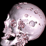
Multiple myeloma is the most common generalized primary neoplasm of the
bones. The cause of this pathology is the monoclonal proliferation of
plasma B cells, resulting in marrow infiltration. Multiple osteolytic
lesions throughout the skeleton are characteristic for multiple myeloma
in radiological examinations.
Material and Methods: This analysis involved twenty-two patients with multiple myeloma, hospitalized in the period between the year 2009 and 2014 in the Hematologic Department, examined with diagnostic imaging techniques.
Results: In all cases, standard X-ray examinations were performed to visualize lytic areas. CT and MRI examinations were carried out when plain X-rays showed no abnormalities in symptomatic areas of the skeleton and to examine parts of the skeleton which cannot be accurately visualized with this method (ribs, sternum). MR imaging was used in twelve patients with neurological symptoms and in two cases it showed cord compression by multiple myeloma infiltration.
Conclusions: The preferred initial imaging method for the diagnosis and staging of multiple myeloma remains the standard X-ray examination. Although it is a conventional examination, it has its limitations. CT should be used to explain ambiguous findings in plain X-rays especially in parts of the skeleton that are difficult to visualize in the standard method, such as ribs, sternum and scapulae. CT and MRI examinations can accurately show the presence and extent of associated soft tissue changes and are useful in the planning of radiotherapy. MRI is the technique of choice for explaining reasons of neurological symptoms suggestive of cord compression. It provides accurate assessment of the level and extent of cord or nerve root compression, size of the tumor and the degree of meningeal infiltration.
Keywords: Magnetic Resonance Imaging, Multiple Myeloma, Tomography Scanners, X-Ray Computed
No comments:
Post a Comment